Methods
Synthesis Laboratory
Schlenk MethodThe Synthesis Laboratory is equipped with a Schlenk line for synthesis of nanomaterials under inert atmospheres. The Schlenk line consists of a dual manifold of several ports. Our setup allows the removal (by vacuum) of the existing atmosphere and the direct incorporation of the inert phase (N2) into the reaction flask. A liquid nitrogen trap and oil bubbler are installed to maintain the pressure and contamination of the system at optimal conditions. High-temperature reactions are also possible by adapting reflux systems and heating mantles to sustain constant reaction volume and temperatures up to 450 °C. | 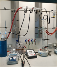 Schlenk Line @biointerfaces lab |
Autoclave reactor methodSteel autoclave reactor (Huanyu 100 mL) with a chemically inert PTFE inner line, allows synthesis at high pressure (up to 30 bar). Together with the natural convection oven (Memmert UM-600), reactions can be performed up to 200 °C. Control of temperature and solution vapor pressure within the reactor enables fine-tuning of the crystal phase and geometries of the desired materials. | 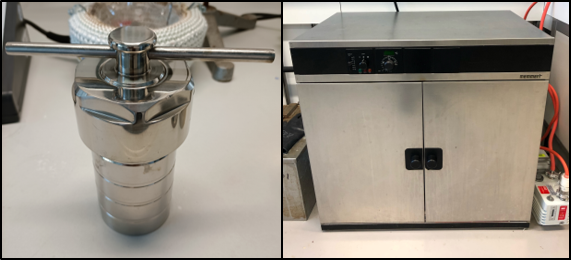 |
Particle separationSeparation of synthesized nanoparticles can be achieved based on their sedimentation coefficient during centrifugation steps. We have two centrifuges to separate larger colloidal particles (ThermoScientific Megafuge, up to 5000 RCF) and for smaller particle sizes (IKA G-L S000, up to 12000 RCF). | 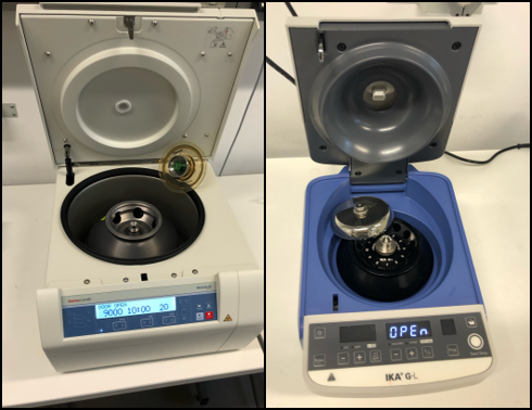 |
Particle dispersionPreparation of nanoparticle suspensions requires proper dispersion of the samples in specific solvents. The Synthesis Laboratory uses a temperature-controlled sonication bath and probe to induce the dispersion of nanomaterials by thermal and/or sonic control of surface interaction between nanoparticles, avoiding the agglomeration of the material due to e.g., van der Waals interactions. | 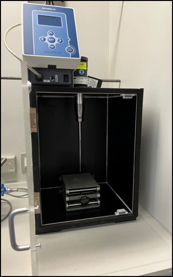 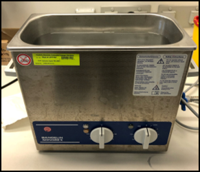 Sonication devices @biointerfaces lab |
Water purification systemReaction synthesis of nanomaterials demand high purity level of solvents and reagents; therefore, the Synthesis Laboratory counts with a Milli-Q Direct 5Q UV water purification unit to assure the use of water type I (18MΩ·cm) by reverse osmosis purification. | 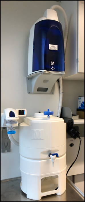 |
Chemical reagent preparationFor preparation of chemical reagents the laboratory is equipped with an analytical scale (VWR LAG254i-M precision up to 0.003g) a shaking unit (IKA Rotator) and a pH-Meter (Fisher-Scientific accumet AE150 pH 0 – 14 / T° 0-90). | 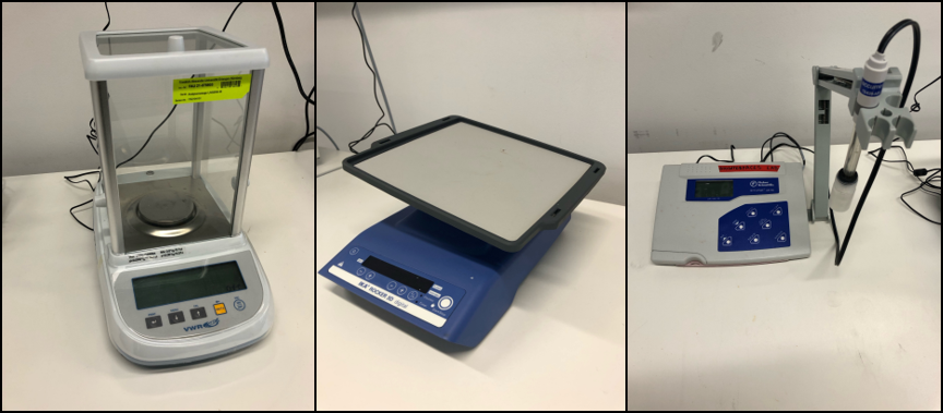 |
Imaging Facility
Inverted fluorescent microscopeInverted fluorescent microscope (Olympus IX7) with ORCA FLASH high speed camera for Calcium imaging and real time cell imaging. Equipped with DAPI, FURA2, TexasRed and ALexa633 filters for multichannel imaging. Recorded data are processed using MATLAB and Adobe image processing applications. | 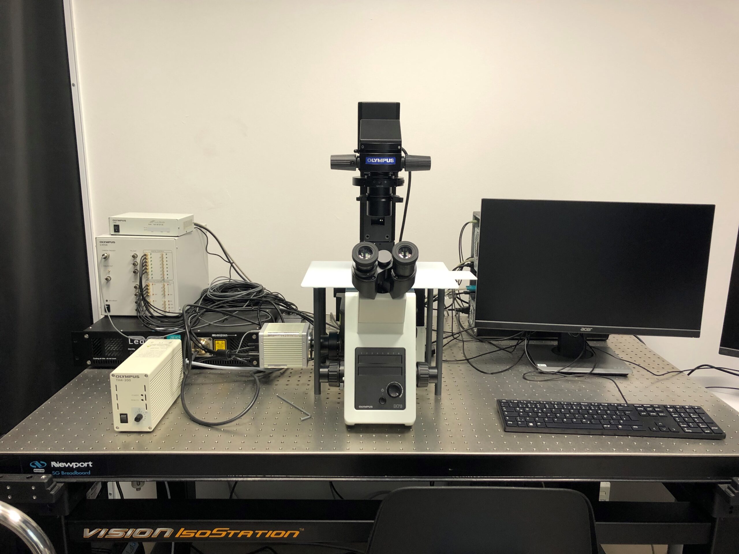 |
| Atomic Force Microscope Asylum Research Cypher S AFM for surface visualization at nanoscale with atomic-level resolution. It allows for accurate characterization of topography, mechanical properties, and surface interactions without requiring complex sample preparation. | 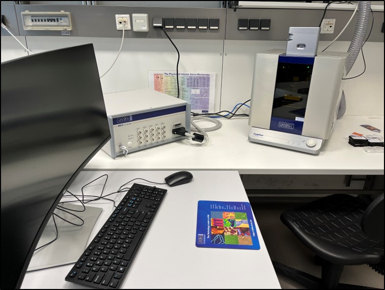 Atomic Force Microscope @biointerfaces lab |
Stimulation Suites
| Custom made magnetic driving electronics for generating aletnating magnetic fields, applicable for magnetic characterisation, in vitro and (rodent) in vivo stimulation: | |
Magnetomechanical suite0-30 Hz and 1-50 mT Magnetothermal suite50kHz- 650 kHz and 10-70 mT Magnetoelectric suiteAC 0-30 Hz and 1-50 mT+ DC 110-230 mT | 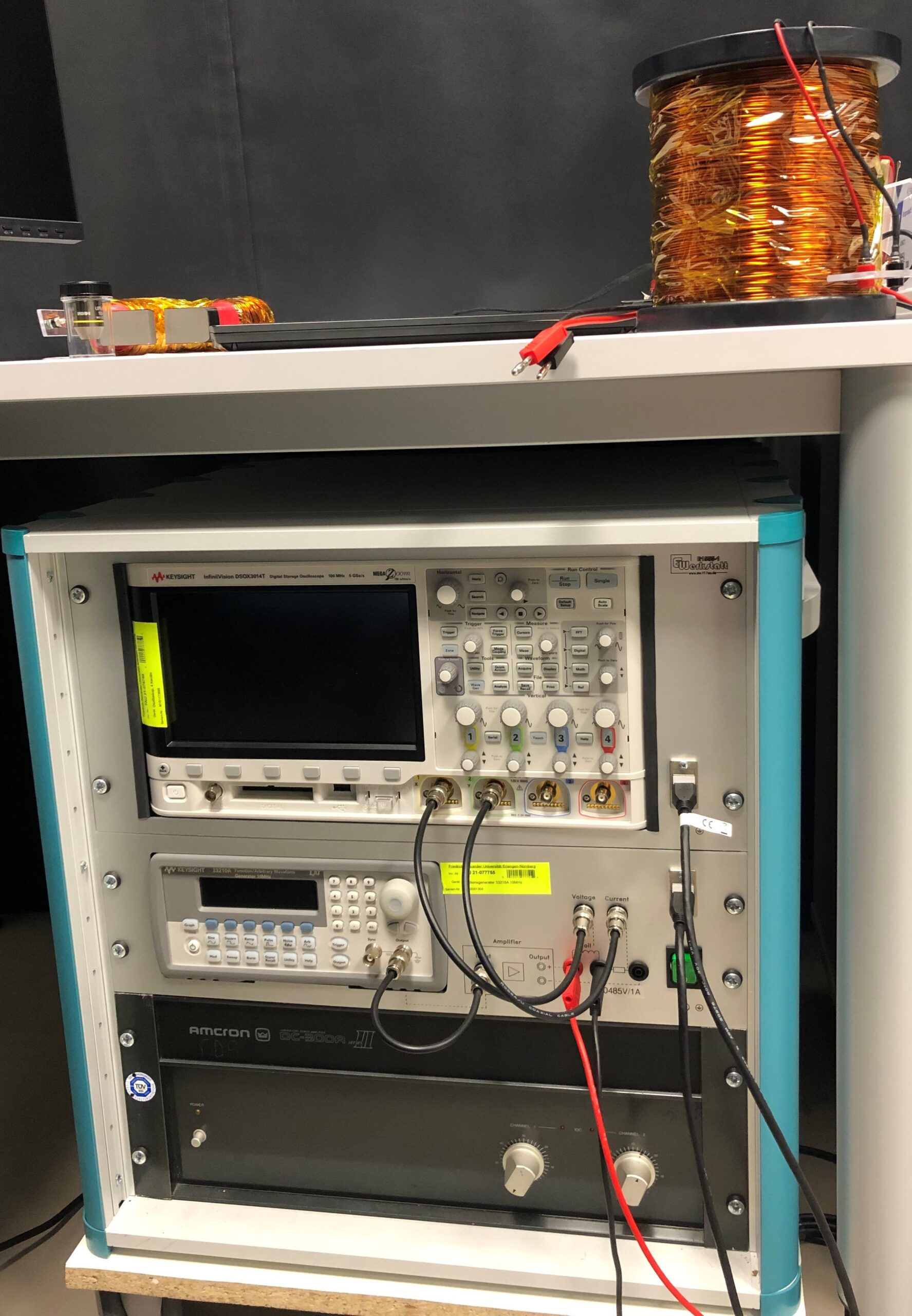 |
Patch Clamp systemElectrophysiological measurements are conducted using a Port-a-Patch patch clamp system (provided by Prof. Andrea Büttner). | |
Molecular Biological Facility
Tuttnauer AutoclaveThe autoclave is used for preparation of sterile materials and media used in the biotechnological laboratories. | 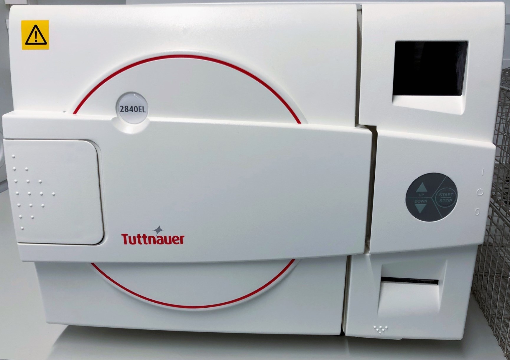 |
Biosafety Cabinet (ENVAIR)All genetic material is handled in the biosafety cabinet. The cabinet provides sterile conditions to work with cell cultures and tissue as well as other biological material. | 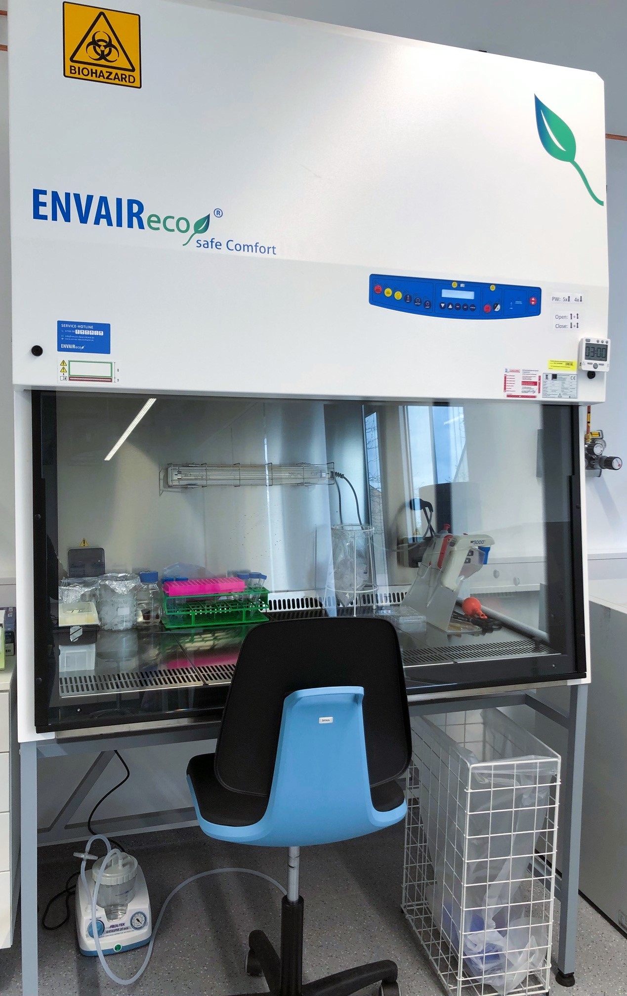 |
Incubators (Memmert)The bio-engineering lab is equipped with two incubators for cultivation of primary neurons and HEK293 cell-lines. | 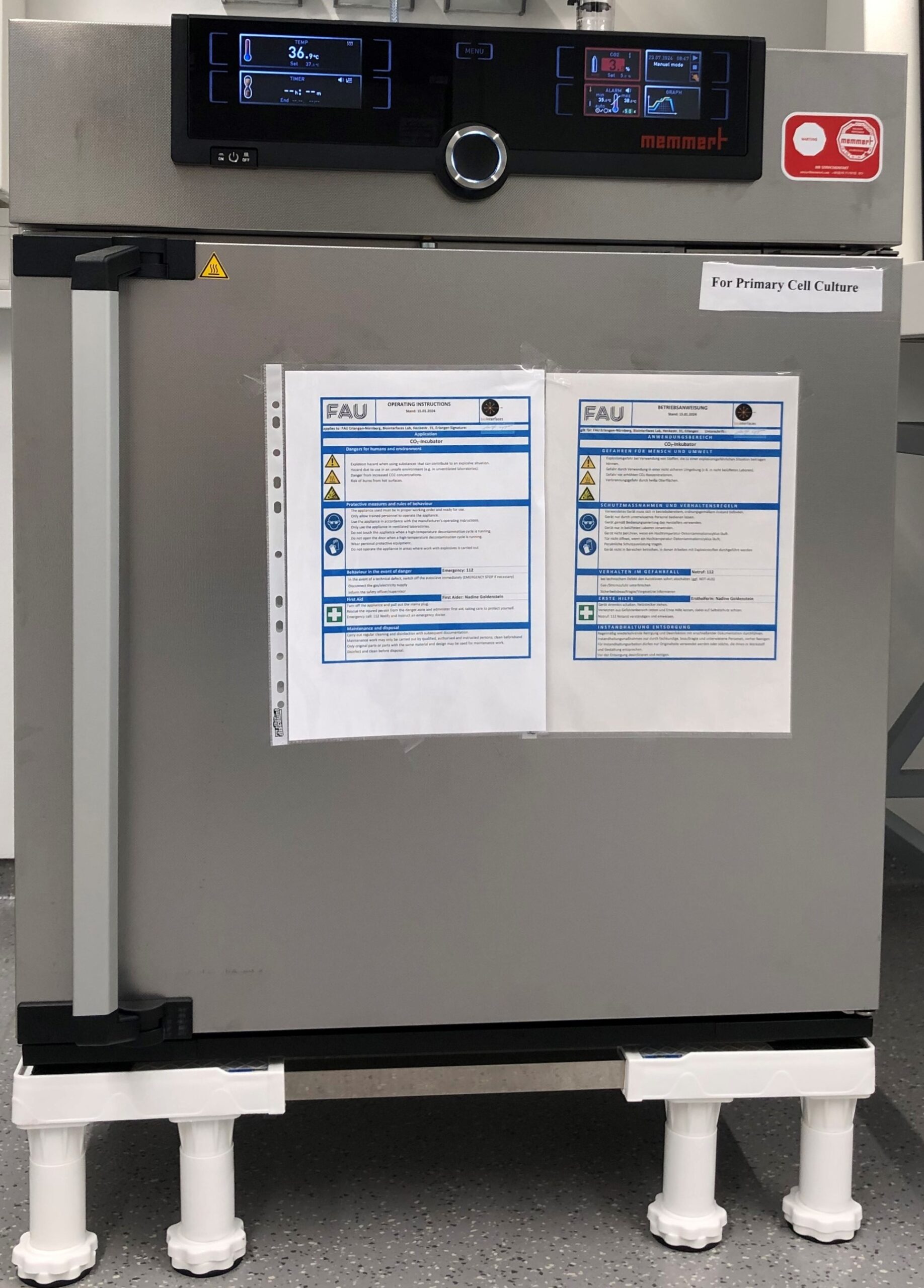 |
Thermocycler (Biometra TOne)PCR can efficiently be performed in our Thermocycler, providing optimal temperature control and easy adjustment of individual protocols. | 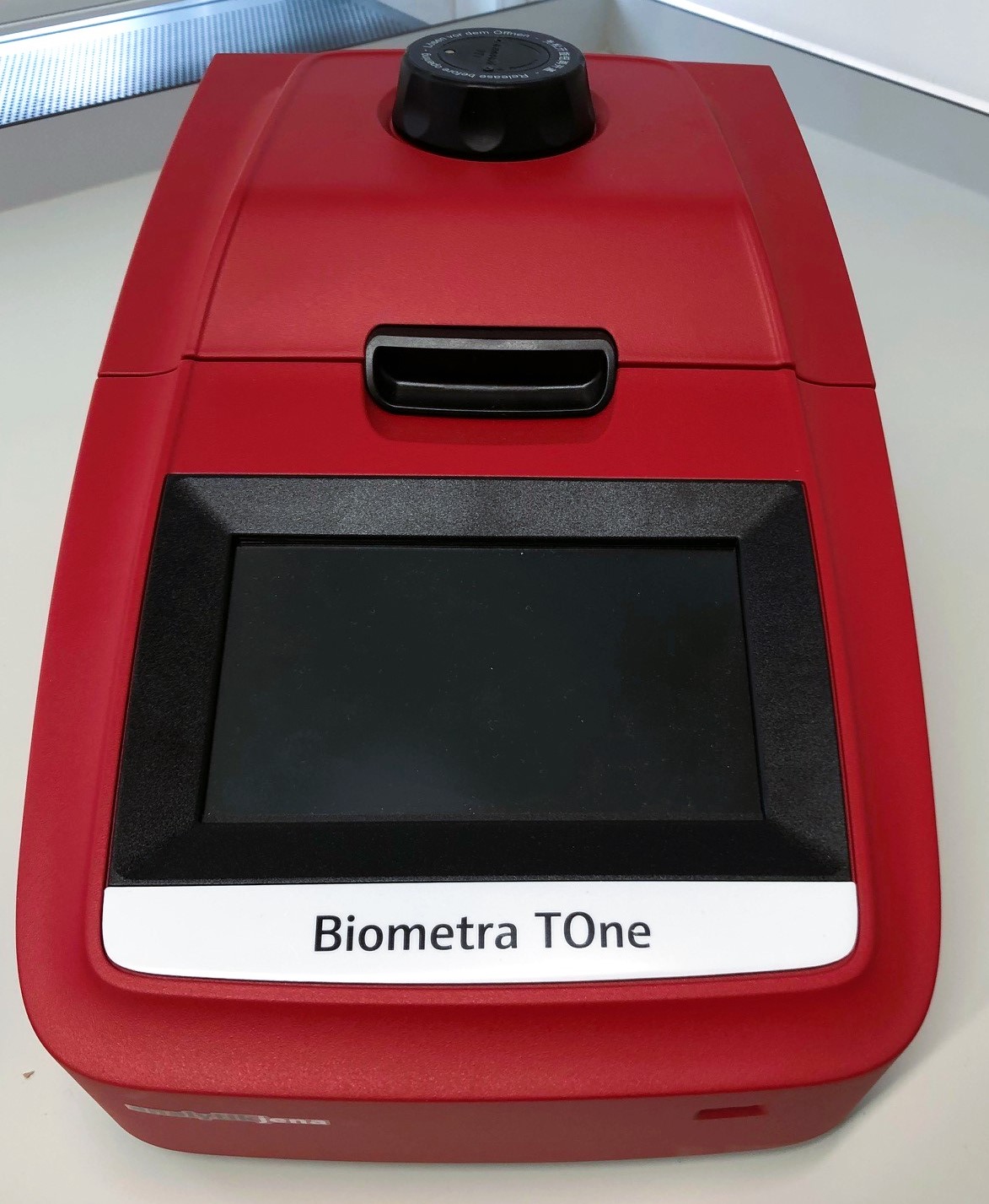 |
Microplate Reader (SpectraMax M2)The multi-detection microplate reader is equipped with dual-monochromators and dual-mode cuvette ports. Detection modalities include absorbance (UV-Vis Abs) and fluorescence intensity (FI). This microvolume spectrophotometer is used to quantify and assess DNA. | 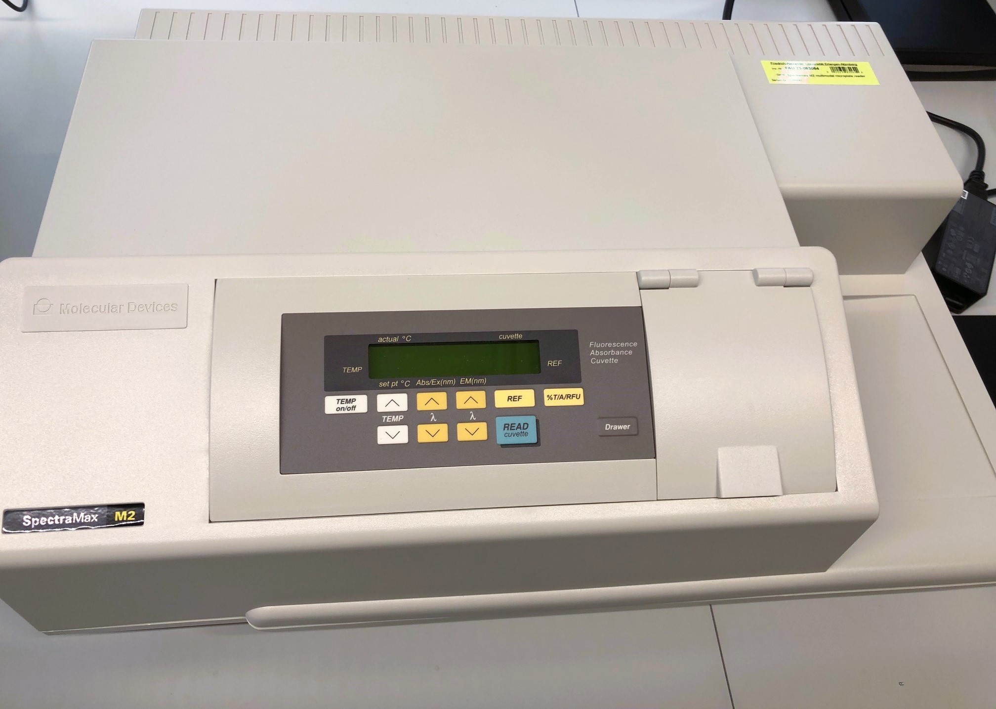 |
Transmitted light microscopeImaging with this microscope is performed during cell culture maintenance and quality control of growth. | 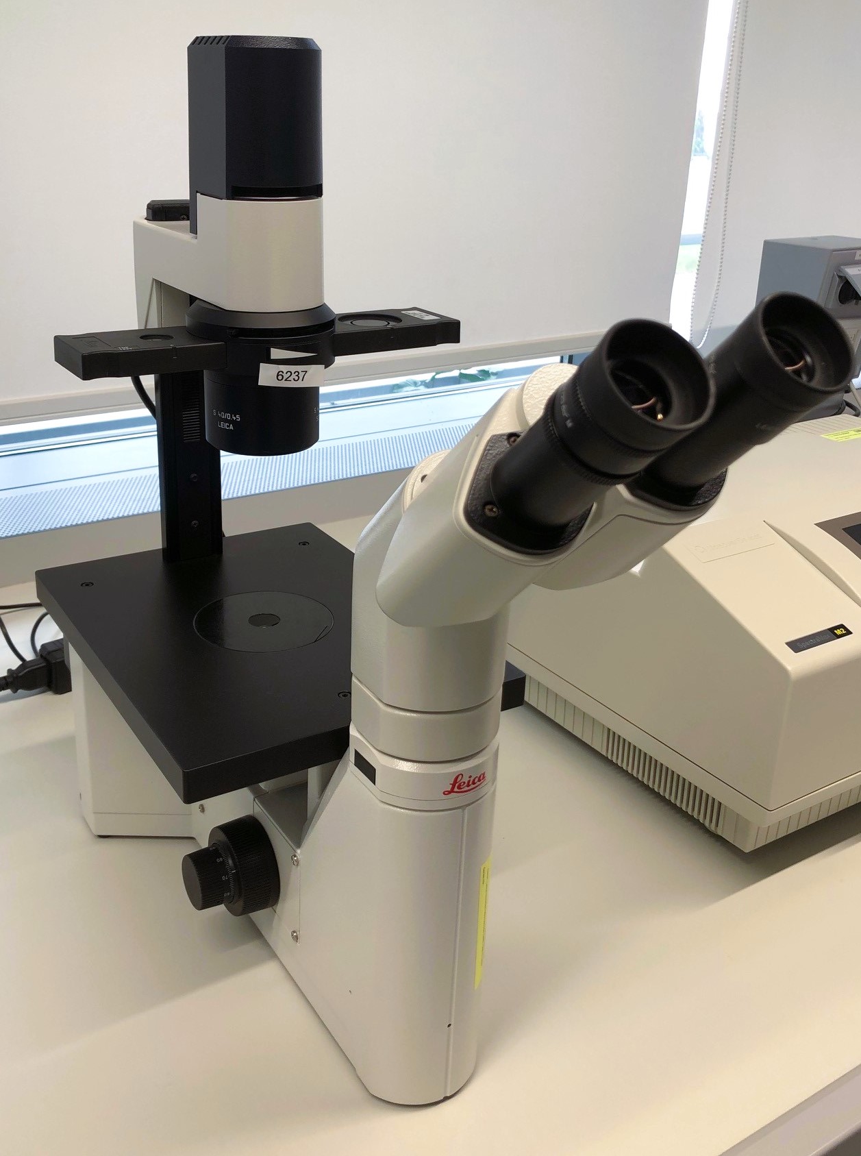 |